
 +86-0755-86131192
 2025-09-10
The arch of the foot is a core structure for human walking, standing, and absorbing impact. Its shape and function directly affect gait stability, lower limb alignment, and overall movement performance. Abnormal arch conditions such as flat feet (collapsed arches) or high arches (excessively elevated arches) can not only lead to plantar fasciitis, Achilles tendonitis, knee pain, and other issues, but may also cause long-term biomechanical imbalances.
Therefore, an objective and accurate assessment of arch condition is a critical prerequisite in rehabilitation medicine, sports science, and personalized orthotic interventions. Traditional methods such as visual inspection or footprint tracing are no longer sufficient to meet the demands of modern precision healthcare. The emergence of foot sole 3D scanners has provided high-precision, quantifiable data support for arch assessment, ushering in a new era of digital foot health evaluation.
Limitations of Traditional Assessment Methods
In clinical practice, arch assessment has long relied on subjective judgment or simple tools, such as foam imprint methods, visual inspection, or angle rulers. These methods have obvious shortcomings: the footprint method is affected by uneven pressure, making it difficult to accurately reflect arch morphology under static or weight-bearing conditions; visual inspection depends on experience, lacks standardized criteria, and is prone to misjudgment; manual measurements suffer from large errors and poor repeatability. More importantly, these methods cannot capture continuous surface data in three-dimensional space, making it difficult to quantify the curvature, height, and dynamic changes of the arch.How 3D Scanning Technology Enables Precise Arch Assessment
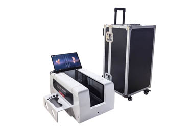
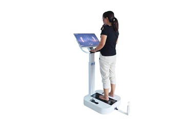
Foot sole 3D scanners use non-contact optical technologies (such as laser scanning or structured light) to complete high-density data acquisition across the entire sole surface within seconds, generating a 3D digital model with millimeter-level accuracy. This technology brings revolutionary improvements to arch assessment:
Accurate Reconstruction of Arch Morphology Under Weight-Bearing Conditions
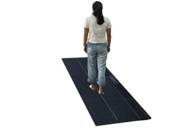
Scanning is typically performed while the subject stands naturally (with single or double foot loading), truly reflecting the biomechanical behavior of the foot under load. Compared to non-weight-bearing conditions, weight-bearing scanning better reveals the degree of arch collapse and avoids "false normal" misdiagnoses.
Precise Quantification of Key Anatomical Parameters
Based on the 3D model, software can automatically identify key anatomical landmarks such as the navicular tuberosity, calcaneal center, and first metatarsal head, and calculate several core metrics:
Arch Height: The vertical distance from the navicular point to the reference plane of the sole, serving as a direct indicator for diagnosing flat feet or high arches.
Longitudinal Arch Index (LAI): Assesses arch type through the ratio of the arch area to the total sole area; a lower value indicates a higher arch.
Arch Curvature Radius: Fits the arc of the medial longitudinal arch and calculates its curvature, reflecting the elasticity and support capacity of the arch.
Visualization Analysis and Longitudinal Tracking
The 3D model can be rotated 360° and zoomed in/out, providing an intuitive display of the arch's three-dimensional shape. Additionally, the system supports multi-timepoint data comparison, facilitating tracking of intervention outcomes (e.g., before and after orthotic insole use) and evaluating rehabilitation progress.
Clinical Application Value
In rehabilitation centers, foot and ankle specialty clinics, and sports training institutions, 3D scanning-based arch assessment is widely applied in:
Pediatric foot development monitoring: Early detection of flat foot tendencies for timely intervention;
Diabetic foot risk screening: Identifying areas of high pressure caused by arch collapse to prevent ulcers;
Sports injury prevention: Customizing supportive insoles for athletes to optimize gait;
Orthotic device design: Providing precise support points and shaping references for custom insoles.Operational Standards and Data Reliability
To ensure measurement accuracy, standardized procedures must be followed: subjects should stand barefoot with evenly distributed weight and minimal body movement; scanning equipment should be regularly calibrated; and software algorithms should be clinically validated. Furthermore, integrating a plantar pressure distribution system allows for further analysis of the relationship between arch function and pressure, achieving dual assessment of both morphology and function.
 Back to list
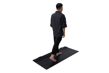


 +86-0755-86131192