
 +86-0755-86131192
 2025-08-11
The following is a professional guide to interpreting foot scanner measurement data, combining 3D modeling with clinical assessment criteria:
I. Key Points for Interpreting Core Data
Arch Morphology Analysis
Normal arch: Medial longitudinal arch angle 113°-130° (angle measured from the lowest point of the calcaneus, head of the talus, and first metatarsal bone).
Flat foot: Medial longitudinal arch angle > 130° and lateral longitudinal arch > 150° (measured in the weight-bearing position).
High-arch foot: Abnormally elevated arch with concentrated plantar pressure points (judgment requires reference to the 3D model color scale).
Foot Type Classification
Egyptian foot: Longest big toe, with linearly decreasing length. Shoes with oblique/square toes.
Greek foot: Second toe longer than big toe. Shoes with pointed toes.
Roman foot: First three toes of similar length. Round-toed shoes/wide-last shoes
Biomechanical abnormality indicators
Hallux valgus: Hallux valgus angle >15° (normal range 0°-10°)
Foot valgus: Abnormally increased calcaneal inclination angle (3D model shows arch collapse)
Example of 2D and 3D report analysis
▲ Typical Scan Report Diagram
Yellow dashed line: Arch height curve (collapse suggests flat feet)
Blue markers: Achilles tendon center and metatarsal key points (used for gait analysis)
III. Key Points for Clinical Diagnosis
Data Validation Standards
A rescan is required if the difference compared to the historical database is greater than 15%.
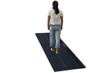
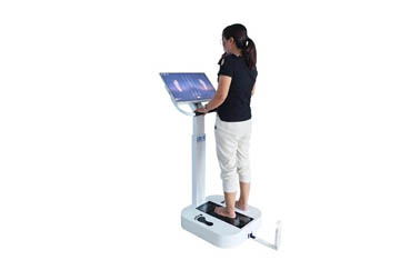
Measurements must be made in the weight-bearing position (sitting data are invalid).
Comprehensive Assessment Recommendations
For suspected flat feet, the talar-first metatarsal angle should be measured in conjunction with an X-ray.
Sports Injury Risk Prediction: A forefoot pressure ratio greater than 40% indicates a need for enhanced cushioning.
Customized insoles require arch width and first metatarsophalangeal joint height data.
Key Note: A single scan cannot directly diagnose foot problems. A comprehensive assessment by a rehabilitation physician, combined with gait analysis and imaging, is required. Before scanning, ensure the soles of the feet are clean and dry, and that weight is evenly distributed when standing.
 Back to list


 +86-0755-86131192