
 +86-0755-86131192
 2025-06-27
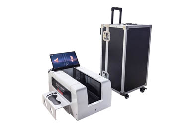
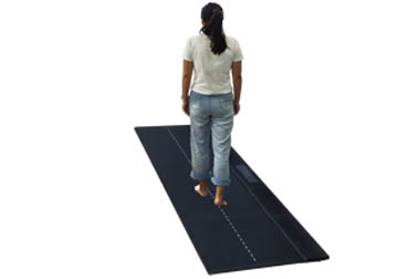
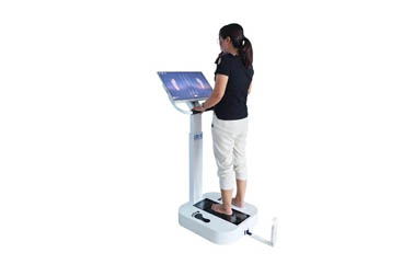
Today, with the focus on the physical development and spinal health of adolescents, "scoliosis" has become a keyword for campus physical examinations and high-incidence screening. In order to detect and intervene early, more and more rehabilitation institutions, posture training centers, and school clinics have begun to introduce a device - a scoliosis 3D scanner. It is radiation-free, contact-free, and has fast output, and is quite popular.
So many people have a question: Can this type of scanner completely replace traditional X-rays? Today's article will dismantle the core logic of this issue and help you understand the true relationship between "high-tech posture screening" and "medical imaging diagnosis".
1. The 3D spine scanner does not rely on "perspective" but on "surface modeling"
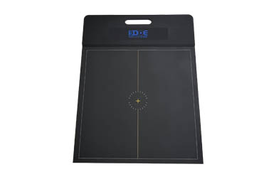
The spine scanner uses the optical scanning principle, usually structured light or laser projection, and uses multiple cameras to collect the surface contour of the human back, including shoulder height, shoulder blade protrusion, spinal curvature direction, pelvic rotation, etc.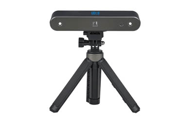
The system analyzes these external morphological features to determine whether there is a suspected scoliosis trend and gives evaluation data such as angle and symmetry. Its advantages are:
No radiation: can be used repeatedly, especially suitable for long-term tracking of children and adolescents
Quick modeling: map building is completed in a few seconds, and reports are issued on site
Strong visualization: patients and family members can intuitively see the changes in body shape
But it should be made clear that it can only "see the surface morphology" and cannot penetrate the skin to identify changes in vertebral structure. This determines that it cannot replace X-rays in medical imaging.
2. X-rays can "penetrate bones", and scanners can only "measure shapes"
Traditional X-ray examinations can directly image vertebral morphology, judge deep structural information such as vertebral rotation, scoliosis angle (such as Cobb angle), and bone changes, which is especially important for diagnosing moderate to severe scoliosis, preoperative and postoperative evaluation, etc.
However, 3D scanners can only provide "surface references":
Suitable for initial screening, trend monitoring, training feedback evaluation
Not suitable for clear diagnosis, pathological classification, and surgical basis
Cannot replace X-rays as formal medical report vouchers
Therefore, in medical clinical scenarios, 3D scanners are more of a screening tool and rehabilitation follow-up tool, rather than a diagnostic terminal.
3. Correct use: combined use rather than substitution
For schools, communities, and rehabilitation institutions, 3D scanners are very suitable as the "first line of defense" - initial screening for abnormal posture, spinal deviation, etc. After positive screening, referral to the hospital for further X-ray examination and diagnosis.
For cases that have been diagnosed and are undergoing training or rehabilitation intervention, 3D scanning can be used for high-frequency tracking, staged evaluation, and efficacy comparison to avoid frequent X-ray exposure and protect children's health.
 Back to list
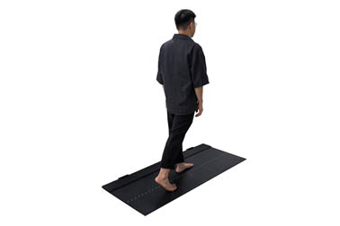


 +86-0755-86131192