
 +86-0755-86131192
 2026-09-09
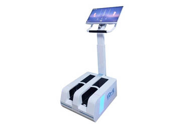
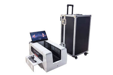
The foot laser 3D modeling scanner mainly uses 3D scanning technologies such as laser and structured light to perform non-contact scanning of the foot surface, obtain data such as the complete 3D geometric shape, length, width, height, angles, and volume of the foot, thereby establishing a 3D digital model of the foot.
The assessments of the foot by the foot laser 3D device mainly include: foot morphology measurement, foot arch morphology assessment, bilateral foot symmetry assessment, and foot dimension measurement.
1. Perform foot 3D morphology assessment through static standing.
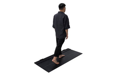
This testing mode is very simple, and it only requires standing barefoot naturally in the scanning area to perform 3D scanning of the foot.
During measurement, it is best to be barefoot, during the measurement process, keep the body naturally relaxed, do not deliberately change the standing posture of the feet, the scanning time is usually very short, and generally the acquisition of the foot 3D data can be completed in a few seconds.
Static 3D scanning mainly analyzes the following aspects:
(1)According to data such as foot length, foot width, instep height, and forefoot width, assess the overall dimensions and 3D morphological characteristics of the foot.
(2)According to data such as the 3D height of the arch area, the plantar contour, and the arch morphology, analyze the arch morphology, which can assist in determining conditions such as a low arch, normal arch, or high arch.
(3)Compare data such as the foot length, foot width, instep height, and arch height of the left and right feet, and assess whether there is obvious asymmetry in the morphology of both feet.
(4)Through 3D data such as heel morphology, forefoot width, instep height, and foot contours, perform an overall assessment of the external morphological characteristics of the foot, providing basic data for footwear selection and subsequent foot intervention.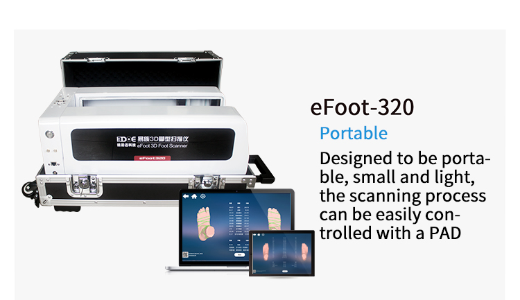
2. Perform foot assessment based on arch morphology.
Foot 3D scanning can obtain the 3D geometric morphology of the plantar and dorsal surfaces of the foot, and perform a more intuitive morphological analysis of the arch area.
During measurement, it is only necessary to stand naturally, keep both feet in a normal weight-bearing state, and the scanner collects foot surface data from multiple angles, so that a complete 3D model of the foot can be formed.
Arch morphology assessment mainly observes the height, width, and plantar contour changes of the arch area.
Through the comparison of arch height, plantar contact contour, and the morphology of the left and right arches, the basic morphological characteristics of the arches can be understood.
Of course, arch morphology is only one part of foot structural assessment, if it is necessary to further determine foot function or disease conditions, other assessment results such as plantar pressure, joint range of motion, and clinical examination also need to be combined for comprehensive assessment.
3. Perform comparative assessment through 3D data of both feet.
When performing foot 3D scanning, complete 3D models of the left and right feet can be obtained simultaneously, and the two foot models can be compared based on their data.
(1)Compare data such as the foot length, foot width, instep height, and arch height of the left and right feet, and determine the differences in the overall dimensions and morphology of both feet.
(2)Through the comparison of foot contour changes in the 3D models, observe whether there are obvious morphological differences between the left and right feet in areas such as the forefoot, arch, and heel.
(3)Through data such as foot length, width, and local height, assist in determining whether there are obvious dimensional inconsistencies between the left and right feet, providing a reference for shoe size selection and separate fitting of the left and right feet.
(4)Compare the foot 3D models obtained from multiple scans, and also observe changes in foot morphology at different times or under different intervention conditions, providing quantitative data for subsequent assessment.
4. Application scenarios.
Through foot 3D scanning data, applications such as foot shape screening, foot arch morphology analysis, shoe size and shoe type selection, foot shape data acquisition before custom insoles / custom shoes, left and right foot morphology comparison, foot development assessment in adolescents, foot shape analysis of athletic populations, and comparison of changes in foot morphology before and after rehabilitation intervention can be carried out.
Note: Foot 3D scanning is mainly a digital assessment tool for the external shape and geometric structure of the foot, mainly reflecting information on the surface morphology and dimensions of the foot, cannot directly reflect plantar pressure, internal bone structure, or muscle and joint function, and cannot independently replace imaging examinations and clinical physical examinations, and can be used for foot shape data acquisition, insole and footwear fitting, and can also be used as a quantitative reference for comparison before and after subsequent intervention.
 Back to list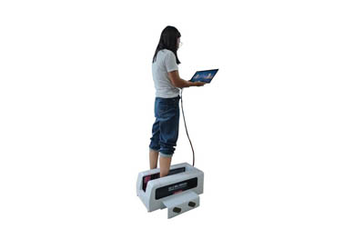


 +86-0755-86131192