
 +86-0755-86131192
 2026-09-03
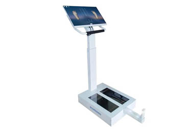
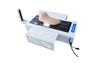
A 3‑D foot scanner is a device that uses high‑precision 3‑D scanning technology to collect data on the shape, dimensions, angles and other parameters of various parts of the human foot while a person stands still.
The 3‑D foot scanner has only one measurement mode: static measurement.
So what data does static 3‑D measurement collect? What are the functions of measuring these data?
Static 3‑D measurement by the foot scanner mainly collects parameters including foot length, width, height, circumference, joint angles, arch and heel morphology, and quantifies the structural and morphological characteristics of foot bones and soft tissues when a person is standing.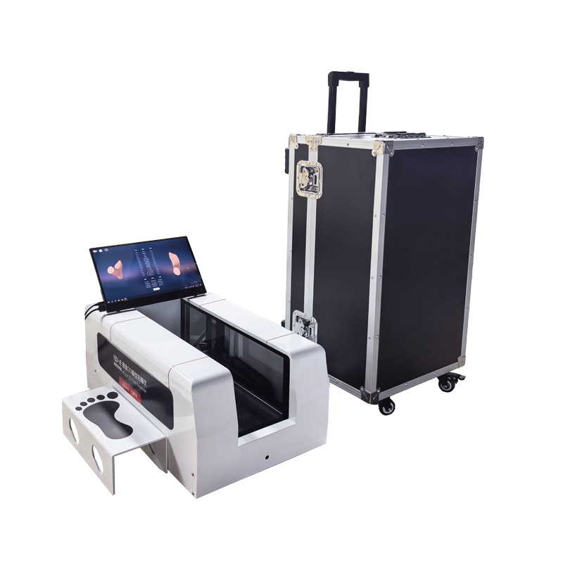
Length data of various foot parts (mm).
The length data of various foot parts refers to the straight‑line distance between key points on the foot; differences in values indicate distinctions in foot bone development and stress‑bearing points; it is used to compare changes in foot morphology before and after orthotic intervention.
Width data of various foot parts (mm).
It includes transverse width parameters at different positions of the foot, representing the transverse width range of each area of the foot contour.
Height data of various foot parts (mm).
This value represents the longitudinal protruding height of each key point from the plantar surface to the instep of the foot.
Circumference data of various foot parts (mm).
This value refers to the contour dimension around different positions of the foot.
Angle data of key foot joints (°).
This value represents the tilt and deflection angles of the toes, metatarsophalangeal joints and heel of the foot.
Classification data of foot arch and heel morphology.
This value represents the classification of foot‑arch height type and heel valgus‑varus morphology.
Abnormalities in angles and morphological classification mean that postural deviation has occurred in the foot bones and joints, and the plantar fascia and foot ligaments are continuously under abnormal traction.
These data can locate structurally weak positions of a person’s foot. Orthotists and rehabilitation therapists can make targeted insole customization and foot correction based on these data, instead of generally adopting universal orthotic products.
They can also be used to detect the sources of some foot‑related symptoms. For example, when the body suffers from plantar soreness after long‑time standing, fascia‑pulling discomfort, abnormal ankle stress or repeated shoe abrasion, the 3‑D scanning data can support targeted analysis.
They can also be applied to screen abnormal foot development. With 3‑D scanning data, professionals can evaluate in advance whether there are developmental abnormalities in the morphology of foot arches, heels and toes. Targeted orthotic intervention can reduce and avoid the risk of subsequent foot sequelae.
Of course, the measured data can only serve as a biomechanical screening tool and cannot be used alone to diagnose diseases. Disease diagnosis requires doctors to combine other professional examinations and make professional judgments.
 Back to list


 +86-0755-86131192